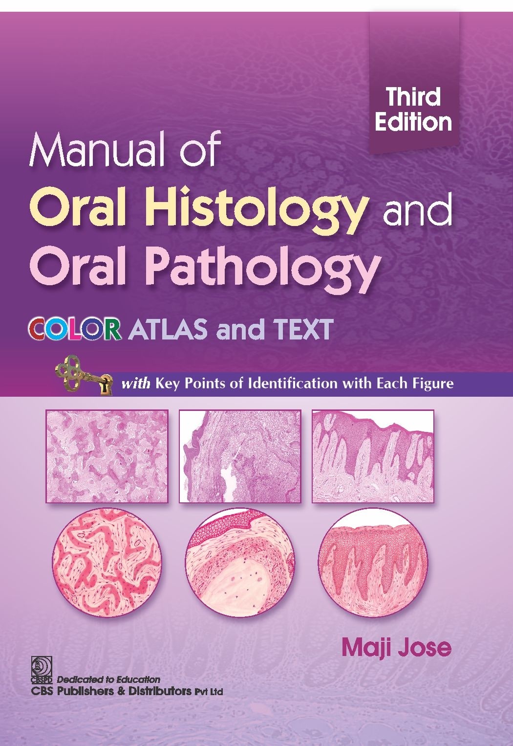
This is the thoroughly revised edition of the book with high quality photomicrographs supporting the drawn diagrams
This popular book is a well-organized guide for oral histology and oral pathology practicals with photomicrographs and neatly hand-drawn diagrams of microscopic structures
All the diagrams are drawn accurately by the author
Precise description of the theoretical aspects in a lucid and simple language
Prominently given are key boxes highlighting the key identification points
Includes an additional section on special stains used in histopathology